Dentistry

Chewing Xylitol Gum Decreases Risk for Ear Infection in Children

Posted on
Share

According to a new study.

Chewing gum containing xylitol may actually prevent ear infections in kids, researchers say.

In a meta-analysis of three Finnish studies, children who chewed gum — or took other products laden with xylitol, including lozenges or syrup — had about a 25% lower risk of developing acute otitis media compared with control interventions, Amir Azarpazhooh, DMD, of the University of Toronto, and colleagues reported in Cochrane Reviews.

“Based on the studies we reviewed, xylitol seems to be a promising alternative to conventional therapies to prevent acute otitis media among healthy children,” they wrote.

Acute otitis media is the most common infection for which kids are treated with antibiotics, which has spurred concerns over antibiotic resistance. So researchers have searched for alternative means of prevention or treatment, not all of which have been successful.

Xylitol, or birch sugar, has been one such alternative. It’s a five-carbon polyol sugar alcohol found in a number of fruits, which has been shown to inhibit the growth and acid production of certain bacteria, particularly S. mutans.

It is for this feature that some dentists recommend it for preventing cavities, the researchers said.

Since a key step in the pathogenesis of otitis media is the colonization of the upper airway with bacteria that move from the nasopharynx to the middle ear via the eustachian tubes, the researchers hypothesized that it may be effective for preventing middle ear infections.

A win – win here. Prevent tooth decay and ear infections.

Hey, a lot better than taking massive amounts of antibiotics.

Share
Cell Phones

Study: Cell Phones Don’t Increase Brain Cancer Risk

Posted on
Share

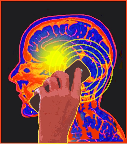
Another study and another finding that there is little worry about your cell phone delivering up brain cancer.

The 5 billion people worldwide who chat away on cell phones shouldn’t worry about an increased risk of brain cancer, new Danish research contends.

One of the largest and longest studies on the subject finds no more brain tumors among people who had cell phones over 17 years than among people who had no cell phones.

Although no one study can rule out harm with absolute certainty, “the risk, if there is one, is extremely low,” said Dr. Ezriel E. Kornel, director of the Neuroscience Institute at Northern Westchester Hospital in Mount Kisco, N.Y.

Previous studies haven’t definitively answered the question of whether cell phone use is harmful: While several studies have found no cause for alarm, a handful did show an upped risk of malignant brain tumors.

Based on the totality of existing evidence, the World Health Organization in May classified cell phones as “possibly carcinogenic to humans,” and placed them in the same category as the pesticide DDT and gasoline engine exhaust.

Experts have been concerned that radio frequency electromagnetic fields sent out by a cell phone held close to the ear could trigger a malignancy.

This new study, led by researchers from the Institute of Cancer Epidemiology in Copenhagen, was a follow-up to an earlier trial that also had found no increased risk in cell phone users. Their latest report is published in the Oct. 20 issue of BMJ.

There are limitations to the study and I am positive the issue will be studied again and again. Will this study satisfy everyone – probably not.

But, good enough for me….at least now.

Share
Diet

Want to Ditch the Twinkies and Other High Caloric Food – Feed Your Brain?

Posted on
Share

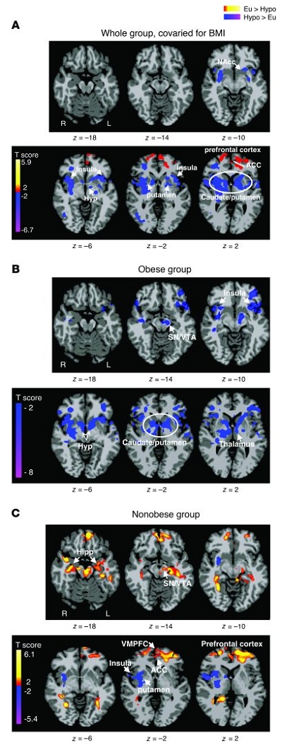
Differences between euglycemic and hypoglycemic conditions.

Axial slices with (A) whole group, covaried for BMI (n = 14), (B) obese group (n = 5), and (C) nonobese group (n = 9) averages, showing brain response to euglycemia compared with mild hypoglycemia across visual cue tasks (threshold of P < 0.05, 2 tailed, FWE whole brain corrected). Red and yellow areas show greater activity during euglycemia, and blue areas indicate greater activity during hypoglycemia. The color scale gives the t value of the functional activity. Eu, euglycemia; Hypo, hypoglycemia; NAcc, nucleus accumbens; Hyp, hypothalamus; VMPFC, ventromedial prefrontal cortex; Hipp, hippocampus; L, left; R, right. MNI coordinates were used to define brain regions.

Yes, according to a new study.

If the brain goes hungry, Twinkies look a lot better, a study led by researchers at Yale University and the University of Southern California has found.

Brain imaging scans show that when glucose levels drop, an area of the brain known to regulate emotions and impulses loses the ability to dampen desire for high-calorie food, according to the study published online September 19 in The Journal of Clinical Investigation.

“Our prefrontal cortex is a sucker for glucose,” said Rajita Sinha, the Foundations Fund Professor of Psychiatry, and professor in the Department of Neurobiology and the Yale Child Study Center, one of the senior authors of the research.

The Yale team manipulated glucose levels intravenously and monitored changes in blood sugar levels while subjects were shown pictures of high-calorie food, low-calorie food and non-food as they underwent fMRI scans.

When glucose levels drop, an area of the brain called the hypothalamus senses the change. Other regions called the insula and striatum associated with reward are activated, inducing a desire to eat, the study found. The most pronounced reaction to reduced glucose levels was seen in the prefrontal cortex. When glucose is lowered, the prefrontal cortex seemed to lose its ability to put the brakes upon increasingly urgent signals to eat generated in the striatum. This weakened response was particularly striking in the obese when shown high-calorie foods.

“This response was quite specific and more dramatic in the presence of high-calorie foods,” Sinha said.

“Our results suggest that obese individuals may have a limited ability to inhibit the impulsive drive to eat, especially when glucose levels drop below normal,” commented Kathleen Page, assistant professor of medicine at the University of Southern California and one of the lead authors of the paper.

A similarly robust response to high-calorie food was also seen in the striatum, which became hyperactive when glucose was reduced. However, the levels of the stress hormone cortisol seemed to play a more significant role than glucose in activating the brain’s reward centers, note the researchers. Sinha suggests that the stress associated with glucose drops may play a key role in activating the striatum.

“The key seems to be eating healthy foods that maintain glucose levels,” Sinha said. “The brain needs its food.”

The abstract for the paper:

Obesity is a worldwide epidemic resulting in part from the ubiquity of high-calorie foods and food images. Whether obese and nonobese individuals regulate their desire to consume high-calorie foods differently is not clear. We set out to investigate the hypothesis that circulating levels of glucose, the primary fuel source for the brain, influence brain regions that regulate the motivation to consume high-calorie foods. Using functional MRI (fMRI) combined with a stepped hyperinsulinemic euglycemic-hypoglycemic clamp and behavioral measures of interest in food, we have shown here that mild hypoglycemia preferentially activates limbic-striatal brain regions in response to food cues to produce a greater desire for high-calorie foods. In contrast, euglycemia preferentially activated the medial prefrontal cortex and resulted in less interest in food stimuli. Indeed, higher circulating glucose levels predicted greater medial prefrontal cortex activation, and this response was absent in obese subjects. These findings demonstrate that circulating glucose modulates neural stimulatory and inhibitory control over food motivation and suggest that this glucose-linked restraining influence is lost in obesity. Strategies that temper postprandial reductions in glucose levels might reduce the risk of overeating, particularly in environments inundated with visual cues of high-calorie foods.

It is common knowledge in running endurance training (e.g. marathon training) that you need to maintain blood glucose levels in order to avoid “negative thoughts” from the brain. Thoughts that will persuade you to stop running.

So, why should this be any different throughout the day or evening when we are not stressing our bodies?

Interesting how obesity plays a role, where an obese person sort of loses their way with regulating their desires for high calorie food.

The entire paper is here. The PDF file is here

Share
Health

Study: Cigarette Smoking Causes More Aterial Damage in Women

Posted on
Share


According to a new study.

The harmful effects of tobacco smoke on atherosclerosis, one of the driving forces of cardiovascular disease, are greater in women than in men.

This result emerges from the large European epidemiological study (Carotid Intima Media thickness and IMT-PROgression as predictors of Vascular Events: the IMPROVE study), funded by EU (Vth Framework Program — Contract n. QLG1-CT-2002-00896). In the IMPROVE study, authors examined 1694 men and 1893 women from Finland, Sweden, Netherlands, France and Italy, and used ultrasound technology to assess the presence of wall thickening and plaques in the carotids, the arteries that bring blood to the brain.

The research shows that the amount tobacco exposure during the entire life significantly correlates with the thickness of carotid arterial walls (an index of atherosclerosis) in both genders. However, the impact is more than doubled in women than in men. Similarly, the effect of the number of cigarettes smoked per day on the progression of the disease over time is more than five-fold in women than in men. These associations are independent from other factors that may affect atherosclerosis, such as age, blood pressure, cholesterol level, obesity and social class.

Elena Tremoli, Professor of Pharmacology at the University of Milan, Italy, and scientific director of the Monzino Cardiology Centre in Milan who led the study, says: “This is a particular relevant finding, especially in view of the fact that educational campaigns carried out in the last years have been less successful in reducing the number of smokers in women than in men.”

According to WHO, while in most European countries a significant proportion of men has quit smoking, in many countries (e.g. Italy and Finland) the percentage of women smokers remained roughly constant in the last three decades, whereas in others (France, Spain) it even increased.

“The reasons for the stronger effect of tobacco smoke on women’s arteries are still unknown, but some hints may come from the complex interplay between smoke, inflammation and atherosclerosis,” says Prof. Tremoli.

Researchers found that other factors, besides smoking, have a differential effect on the arteries of men and women. One of these is education, a well known index of social class: while men who have studied less showed a greater thickening of arterial walls than those who have studied more, the same was not true for women. Similarly, women, in contrast with men, seem to be protected against the harmful effects of systemic inflammation. Indeed, in the IMPROVE study, the relation between arterial wall thickening and the levels of C-Reactive Protein (CRP) and white blood cells (WBC) counts, two indexes of inflammation, is very strong in men, but absent in women.

“It is important to mention, however, that, when women smoke they lose their protection against the harmful effect of inflammation. In particular, if we stratify the female population according to smoking habits, we see that in the group of women who smoke, especially in heavy smokers, the relationship between CRP and arterial wall thickening becomes similar to that observed in men,” says Prof. Tremoli.

“We all know that women are ‘naturally’ protected against cardiovascular disease, particularly before menopause, and this has led to less attention of health professionals and researchers in regard to this disease in women.

Women, who for some reason appear to be less vigilant about heart disease, should take this study to their own heart. If you smoke, you have an increased risk of heart disease – more than the men.

So, stop smoking!

Share
Medicine

Poll Watch: Southern and Western United States Have Highest Uninsured Rates

Posted on
Share


According to the latest Gallup Poll.

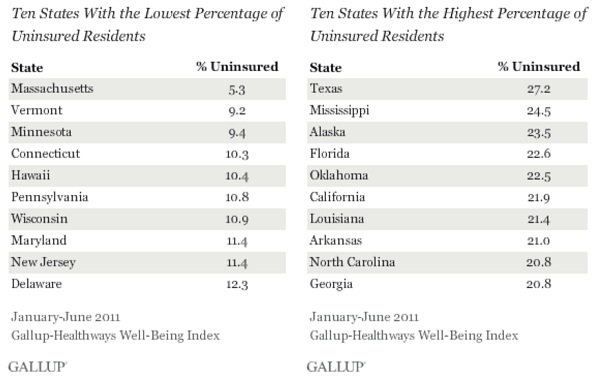
Texas residents continue to be the most likely in the United States to lack health coverage, with 27.2% reporting being uninsured in the first half of 2011. At the other end of the spectrum is Massachusetts, where health insurance is required and 5.3% of residents lack coverage. These two states have represented the upper and lower bounds of uninsured rates since Gallup and Healthways started tracking coverage in 2008.

These results are based on 177,237 interviews conducted daily from January through June 2011 as part of the Gallup-Healthways Well-Being Index. An average of 16.8% of all American adults were uninsured in the first half of 2011, similar to the 16.4% in 2010. This percentage, however, has been edging up each year since 2008, at which time 14.8% of adults were uninsured. The percentage of uninsured residents in all states so far in 2011 is on par with 2010, but in most states remains higher than in 2008.

And, the Southern and Western United States have the highest uninsured rates.

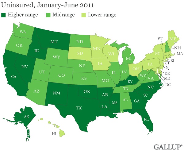
States in the South and West continue to have higher numbers of uninsured adults than do those in the Northeast — consistent with what Gallup found in 2008, 2009, and 2010. Eight of the 10 states with the highest uninsured rates in the country are in the South and the other two — California and Alaska — are in the West.

Texas, California, and Florida — all three of which have an uninsured rate higher than 20% — have disproportionately large Hispanic populations, the demographic group Gallup finds to be the most likely to be uninsured.

Uninsured rates are lower in the Northeast, with 7 of 10 states with the fewest uninsured residents located there, as in past years.

The chart:

So, what does this mean?

President Obama’s Affordable Care Act has had little effect on people being insured or not. With the states struggling with budgets, it is difficult to see how many more people can be provided insurance without affecting the people that already have coverage.

Uninsured rates across states in 2011 appear to be relatively stable so far compared with 2010, but remain higher than in 2008. This could be seen as good news at a time when states are grappling with deep budget cuts and the implementation of new regulations and programs required under the Affordable Care Act. However, more than 10% of adults lack healthcare coverage in almost all states, with more than 15% going uninsured in 29 states. The fate of uninsured rates in America remains precarious as numerous states are challenging the legality of the new healthcare law in court.

Share