Diet

Want to Ditch the Twinkies and Other High Caloric Food – Feed Your Brain?

Posted on
Share

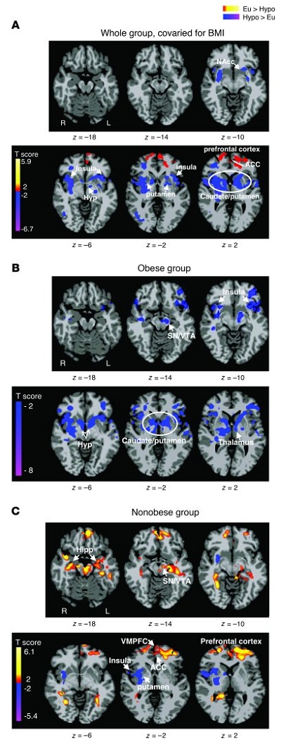
Differences between euglycemic and hypoglycemic conditions.

Axial slices with (A) whole group, covaried for BMI (n = 14), (B) obese group (n = 5), and (C) nonobese group (n = 9) averages, showing brain response to euglycemia compared with mild hypoglycemia across visual cue tasks (threshold of P < 0.05, 2 tailed, FWE whole brain corrected). Red and yellow areas show greater activity during euglycemia, and blue areas indicate greater activity during hypoglycemia. The color scale gives the t value of the functional activity. Eu, euglycemia; Hypo, hypoglycemia; NAcc, nucleus accumbens; Hyp, hypothalamus; VMPFC, ventromedial prefrontal cortex; Hipp, hippocampus; L, left; R, right. MNI coordinates were used to define brain regions.

Yes, according to a new study.

If the brain goes hungry, Twinkies look a lot better, a study led by researchers at Yale University and the University of Southern California has found.

Brain imaging scans show that when glucose levels drop, an area of the brain known to regulate emotions and impulses loses the ability to dampen desire for high-calorie food, according to the study published online September 19 in The Journal of Clinical Investigation.

“Our prefrontal cortex is a sucker for glucose,” said Rajita Sinha, the Foundations Fund Professor of Psychiatry, and professor in the Department of Neurobiology and the Yale Child Study Center, one of the senior authors of the research.

The Yale team manipulated glucose levels intravenously and monitored changes in blood sugar levels while subjects were shown pictures of high-calorie food, low-calorie food and non-food as they underwent fMRI scans.

When glucose levels drop, an area of the brain called the hypothalamus senses the change. Other regions called the insula and striatum associated with reward are activated, inducing a desire to eat, the study found. The most pronounced reaction to reduced glucose levels was seen in the prefrontal cortex. When glucose is lowered, the prefrontal cortex seemed to lose its ability to put the brakes upon increasingly urgent signals to eat generated in the striatum. This weakened response was particularly striking in the obese when shown high-calorie foods.

“This response was quite specific and more dramatic in the presence of high-calorie foods,” Sinha said.

“Our results suggest that obese individuals may have a limited ability to inhibit the impulsive drive to eat, especially when glucose levels drop below normal,” commented Kathleen Page, assistant professor of medicine at the University of Southern California and one of the lead authors of the paper.

A similarly robust response to high-calorie food was also seen in the striatum, which became hyperactive when glucose was reduced. However, the levels of the stress hormone cortisol seemed to play a more significant role than glucose in activating the brain’s reward centers, note the researchers. Sinha suggests that the stress associated with glucose drops may play a key role in activating the striatum.

“The key seems to be eating healthy foods that maintain glucose levels,” Sinha said. “The brain needs its food.”

The abstract for the paper:

Obesity is a worldwide epidemic resulting in part from the ubiquity of high-calorie foods and food images. Whether obese and nonobese individuals regulate their desire to consume high-calorie foods differently is not clear. We set out to investigate the hypothesis that circulating levels of glucose, the primary fuel source for the brain, influence brain regions that regulate the motivation to consume high-calorie foods. Using functional MRI (fMRI) combined with a stepped hyperinsulinemic euglycemic-hypoglycemic clamp and behavioral measures of interest in food, we have shown here that mild hypoglycemia preferentially activates limbic-striatal brain regions in response to food cues to produce a greater desire for high-calorie foods. In contrast, euglycemia preferentially activated the medial prefrontal cortex and resulted in less interest in food stimuli. Indeed, higher circulating glucose levels predicted greater medial prefrontal cortex activation, and this response was absent in obese subjects. These findings demonstrate that circulating glucose modulates neural stimulatory and inhibitory control over food motivation and suggest that this glucose-linked restraining influence is lost in obesity. Strategies that temper postprandial reductions in glucose levels might reduce the risk of overeating, particularly in environments inundated with visual cues of high-calorie foods.

It is common knowledge in running endurance training (e.g. marathon training) that you need to maintain blood glucose levels in order to avoid “negative thoughts” from the brain. Thoughts that will persuade you to stop running.

So, why should this be any different throughout the day or evening when we are not stressing our bodies?

Interesting how obesity plays a role, where an obese person sort of loses their way with regulating their desires for high calorie food.

The entire paper is here. The PDF file is here

Share
Alzheimer's Disease

Does Diabetes Increase Alzheimer’s Disease Risk?

Posted on
Share

Yes, and other forms of dementia as well.

People with diabetes are at increased risk of having a heart attack or stroke at an early age, but that’s not the only worry. Diabetes appears to dramatically increase a person’s risk of developing Alzheimer’s disease or other types of dementia later in life, according to a new study conducted in Japan.

In the study, which included more than 1,000 men and women over age 60, researchers found that people with diabetes were twice as likely as the other study participants to develop Alzheimer’s disease within 15 years. They were also 1.75 times more likely to develop dementia of any kind.

“It’s really important for the [public’s] health to understand that diabetes is a significant risk factor for all of these types of dementia,” says Rachel Whitmer, Ph.D., an epidemiologist in the research division of Kaiser Permanente Northern California, a nonprofit health-care organization based in Oakland, California.

Whitmer, who studies risk factors for Alzheimer’s but wasn’t involved in the new research, stresses that many questions remain about the link between diabetes and dementia. The new study was “well done” and provides “really good evidence that people with diabetes are at greater risk,” she says, “but we really need to look at other studies to find out why.”

More studies are needed and it makes sense that the longer you live, the more of a chance you will develop some sort of dementia. But, it also makes sense that via diet and exercise, you should avoid diabetes in the first place.

Diabetes could contribute to dementia in several ways, which researchers are still sorting out. Insulin resistance, which causes high blood sugar and in some cases leads to type 2 diabetes, may interfere with the body’s ability to break down a protein (amyloid) that forms brain plaques that have been linked to Alzheimer’s. High blood sugar (glucose) also produces certain oxygen-containing molecules that can damage cells, in a process known as oxidative stress.

In addition, high blood sugar — along with high cholesterol — plays a role in the hardening and narrowing of arteries in the brain. This condition, known as atherosclerosis, can bring about vascular dementia, which occurs when artery blockages (including strokes) kill brain tissue.

“Having high glucose is a stressor to the nervous system and to the blood vessels,” says David Geldmacher, M.D., a professor of neurology at the University of Alabama at Birmingham. “The emerging information on Alzheimer’s disease and glucose shows us that we do need to remain vigilant on blood sugar levels as we get older.”

Share
Diet

The Deadly Link Between High Dietary Salt Intake and Obesity

Posted on
Share


New research indicates abnormal activation of a protein may explain the deadly combination of salt intake and obesity.

Dietary salt intake and obesity are two important risk factors in the development of high blood pressure. Each packs its own punch, but when combined, they deliver more damage to the heart and kidneys than the sum of their individual contributions. Discovering the molecular mechanisms behind this lethal synergy has presented a challenge to scientists, but research led by Toshiro Fujita, MD, professor and chairman of the Department of Internal Medicine and chief of the Department of Nephrology and Endocrinology at the University of Tokyo, suggests that high dietary salt intake and obesity work together to trigger an abnormal activation of a cellular protein called Rac1.

According to Dr. Fujita, the team’s findings carry important implications for the treatment of hypertension. “Our data indicate that the Rac1-mediated pathway in the kidneys can be an alternative therapeutic target for salt-sensitive hypertension and salt-mediated kidney injury,” he said. “Based upon our results, we can speculate that Rac1 in the kidneys regulates salt susceptibility of blood pressure, and that Rac1 inhibitors, as well as MR antagonists, may be effective in the treatment of salt-sensitive hypertension.”

In the meantime though, if you are obese lose the weight and ingest ONLY the appropriate amount of dietary salt.

But, this is good news for patients with persistent high blood pressure and may offer a pathway to mediate the combination effects of dietary salt and obesity.

Share
Health

CDC: Successful Anti-Smoking Efforts Resulting in Less Lung Cancer?

Posted on
Share

Former smoker Debie Austin warns viewers about the dangers of smoking.

Yes, according to a new report.

New diagnoses of lung cancer fell from 2006 to 2008 among Americans of both sexes — the first time in decades that the incidence in women has declined, and probably because of successful anti-smoking efforts, the CDC reported.

Nationwide, the reductions in incidence amounted to 2.2% among women from 2006 to 2008 and 2.9% among men from 2005 to 2008, according to data from two CDC registries appearing Thursday in Morbidity and Mortality Weekly Report.

Accompanying and probably explaining these trends were declines in smoking prevalence and increases in so-called quit ratios, seen in responses to the CDC’s Behavioral Risk Factor Surveillance System survey, the report indicated.

Quit ratios are the number of “former smokers” (those responding that they did not currently smoke but had previously smoked at least 100 cigarettes during their lifetimes) divided by the total of current and former smokers.

States with higher quit ratios — which the authors suggested were the result of media campaigns, tobacco tax hikes, and other anti-smoking programs — had greater decreases in new lung cancer diagnoses (r=-0.55 for men, -0.33 for women, both P<0.001).

During this period of time, some states also regulated where smoking could take place (due to second hand smoke). With smoking no longer being socially acceptable inside a residence or a restaurant, some of the glamor and more of the pain in the ass type of habit meme becomes more common place.

Graphic videos like the one above may have had some effect.

But, whatever, I will accept less lung cancer as a worthy societal goal.

Although the authors offered no systematic data to link these trends with state-level tobacco control efforts, they argued that these were likely responsible, at least in part.

They pointed to California as a state that has “invest[ed] more fully in these programs” and has also seen “decreases in youth and adult smoking prevalence, decreases in lung cancer, and significant healthcare savings.”

Share
Health

Virtual Colonoscopy to Become the Standard to Detect Colon Cancer?

Posted on
Share

Virtual colonoscopy images

Well, perhaps or not.

Whether the most technologically advanced way to check for colon cancer will become the standard screening method of the future does not appear to be a slam-dunk.

The method, known as virtual colonoscopy, combines X-ray and computer technology to create three-dimensional views of the full length of the colon, the large intestine. It allows doctors to look for polyps, or pre-cancerous growths, or other signs of cancer or other intestinal disease. According to the U.S. National Cancer Institute, virtual colonoscopy can be done with computed tomography (called a CT or CAT scan) or with magnetic resonance imaging (MRI).

Colon cancer is one of the few types of preventable cancer, with doctors able to find and remove pre-cancerous polyps in the colon before cancer can develop. The current “gold standard” procedure for colon cancer screening, however, is colonoscopy, a time-consuming procedure for which preparation is unpleasant and sedation is necessary.

Perhaps because of this, only half of all people older than 50 have gotten this potentially life-saving test for colon cancer, according to the U.S. Centers for Disease Control and Prevention.

Doctors who tout the virtual form of colonoscopy argue that it takes less time and does not require sedation and is a more comfortable procedure for those having it.

Yet others contend that its drawbacks far outweigh its benefits.

“It’s a test that has a tremendous number of questions still yet to be answered,” said Dr. David A. Johnson, chief of gastroenterology at Eastern Virginia Medical School, past president of the American College of Gastroenterology and co-author of the group’s guidelines for colon cancer screening.

Virtual colonoscopy, however, has advanced far enough that it’s now recommended as a frontline screening test by the American Cancer Society and as an alternative to regular colonoscopy by the American College of Gastroenterology.

I postponed my first test a few more years than I should have, but I have to say it was not a bad procedure. Now, the colon prep is a different sotry since you take powerful laxatives and the abdominal cramping is not fun.

But, you have to do the same prep in cleaning out your colon before the virtual colonoscopy anyway.

If the virtual test allows more patients to receive “some” colon cancer screening, then this is a good thing. I am positive the virtual tests will only improve with the technology and time.

Researchers also are working to make virtual colonoscopy even better, Yee said. New computerized, post-processing techniques are being developed to improve the test’s accuracy and lower the radiation dose, and doctors are working to develop a form of the test for which the patient would not need to take laxatives in advance.

At the moment, though, the American College of Gastroenterology still would prefer that people have a conventional colonoscopy done every 10 years because, as Johnson said, it’s a better test that allows doctors to immediately remove any polyps that are found.

However, if the drawbacks of a normal colonoscopy are enough to dissuade a person from undergoing colon cancer screening, then they should consider virtual colonoscopy as an alternative test that they should have every five years, according to ACG guidelines.

Share